The Hidden Cellular Survival Mechanism That Drives Tissue Regeneration and Cancer Recurrence

For over half a century, biologists have been puzzled by a phenomenon known as compensatory proliferation: the process by which damaged epithelial tissues, such as skin or organ linings, orchestrate a precise and rapid recovery following severe trauma. While the observation that tissues can "rebuild" themselves has been well-documented since the 1970s, the precise molecular switches that govern this miraculous recovery have remained elusive. Now, a groundbreaking study published in the journal Nature Communications by researchers at the Weizmann Institute of Science has unveiled a previously unknown cellular survival mechanism that not only explains how tissues regenerate but also suggests a dark side: this same mechanism may be the primary culprit behind the recurrence of aggressive, treatment-resistant cancers.
A Historical Perspective on Compensatory Proliferation
The foundation for this research dates back to the 1970s, when developmental biologists first observed that fruit fly larvae (Drosophila melanogaster) possessed an extraordinary capacity to repair their bodies. In these early experiments, researchers exposed larvae to high doses of ionizing radiation. Despite sustaining massive cellular damage to their epithelial layers, the larvae were capable of regenerating fully functional, anatomically correct wings.
This discovery challenged the prevailing view of the time, which held that damaged cells simply died and were replaced by stem cells. Instead, the fly models suggested that the body possesses a sophisticated "emergency protocol" to manage mass cell death. Over the following decades, similar regenerative responses were observed across various species, including mammals, yet the molecular "trigger" that distinguished a cell destined for death from one destined to lead the regeneration process remained a mystery.
The DARE and NARE Discovery
To solve this riddle, a team led by Dr. Tslil Braun in the laboratory of Professor Eli Arama of the Weizmann Institute’s Department of Molecular Genetics utilized advanced genetic labeling and high-resolution imaging. By recreating the classic radiation experiments in fruit flies, the team sought to isolate the specific cells that survive what should be a lethal "self-destruct" signal.
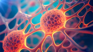
The researchers discovered two distinct populations of cells, which they termed DARE (Death-Associated Recovery) cells and NARE (Non-Death-Associated Recovery) cells. The DARE cells are the primary protagonists of the repair process. These cells initiate the standard apoptosis pathway—a programmed "suicide" mechanism—but then hit a molecular "brake" that halts the process before the cell is destroyed.
"We set out to identify cells that push the self-destruct button but survive anyway," Dr. Braun explained. These DARE cells do more than simply persist; they enter a hyper-proliferative state. Within 48 hours of the initial radiation exposure, these survivors had multiplied rapidly to replenish nearly 50 percent of the damaged tissue.
The NARE cells, by contrast, act as a supporting cast. They do not activate the initiator caspase enzymes and are not triggered by the same initial death signals. However, the study revealed a sophisticated "crosstalk" between these populations. DARE cells secrete growth factors to stimulate NARE cell multiplication, while NARE cells, in turn, release inhibitory signals that prevent the DARE cells from proliferating uncontrollably. This elegant negative-feedback loop ensures that the tissue stops growing once the repair is complete, preventing the development of disorganized tissue masses.
The Molecular Mechanism: Caspases and Motor Proteins
The key to the DARE cells’ survival lies in a protein—a molecular motor—that physically tethers the initiator caspase to the cell membrane. In a normal apoptotic event, initiator caspases trigger a cascade of effector caspases that dismantle the cell’s internal machinery. In DARE cells, this motor protein prevents the initiator caspase from ever reaching the effector phase.
When the research team artificially silenced this motor protein in the lab, the DARE cells failed to stall their death process and subsequently succumbed to the radiation, resulting in a total failure of tissue regeneration. The significance of this finding is profound: the overactivation of this same motor protein has been previously identified in various human cancers. This suggests that cancer cells may be "hijacking" a natural, beneficial repair mechanism to render themselves immortal, effectively cloaking themselves in a survival strategy designed for healthy wound healing.
Implications for Oncology and Treatment Resistance
Perhaps the most alarming discovery from the Arama laboratory involves the "biological legacy" of these cells. When the researchers exposed the regenerated tissue to a second round of radiation, they found that the descendants of DARE cells were seven times more resistant to cell death than the original, unexposed tissue.
This suggests that radiation therapy—intended to kill cancer cells—may inadvertently act as a selective pressure that "trains" surviving cells to become more aggressive and resistant to future interventions. This provides a compelling scientific explanation for why recurrent tumors are often significantly more difficult to treat than the primary, initial malignancy. The tumor is not merely "coming back"; it is returning with an enhanced, evolved survival toolkit.
Future Directions and Clinical Potential
The potential for medical translation is significant. By understanding how these cells escape their "death sentence," clinicians may eventually be able to develop therapeutic agents that block this survival pathway in cancer cells, thereby making them vulnerable to conventional radiation and chemotherapy. Conversely, the ability to safely promote DARE cell activity could lead to revolutionary treatments for severe wounds, burns, or organ damage where rapid, controlled tissue regeneration is required.
Prof. Eli Arama, who holds the Harry Kay Professorial Chair of Cancer Research, emphasizes that while the study was conducted in fruit flies, the fundamental mechanisms of apoptosis and cell signaling are highly conserved across species. "Many cancers originate in epithelial cells that have lost normal growth control," Arama noted. "Our findings pave the way for understanding why such treatments sometimes fail and how they could be improved. We are looking at a system that is essentially a double-edged sword: it is the body’s most effective tool for healing, but in the wrong context, it is the mechanism that allows cancer to thrive."
Broader Scientific Analysis
The study represents a paradigm shift in how cell death is viewed in oncology. For decades, apoptosis was considered a binary "on-off" switch. The existence of DARE cells proves that there is a "paused" state of apoptosis, a biological limbo that allows cells to retain their viability while undergoing the stress of extreme damage.
Independent experts in the field of regenerative medicine have noted that the feedback loop between DARE and NARE cells serves as a vital safeguard. The study provides a rare, high-resolution look at the signaling pathways that maintain homeostasis after trauma. Future studies will likely focus on mapping the specific growth factors exchanged between these cell types in mammalian models, moving the research closer to human clinical trials.
As research continues, the dual nature of this survival mechanism will remain a focal point. Scientists are now tasked with the challenge of finding the "goldilocks" zone—where tissue repair is stimulated in healthy patients, while the "survival brake" is permanently disabled in patients battling metastatic disease. By deciphering the molecular vocabulary that cells use to decide whether to die or multiply, the Weizmann team has opened a new front in the battle against cancer and a promising pathway for regenerative medicine.
Related posts: